Start with the function that needs to be modelled
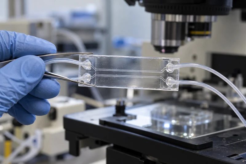
An organ chip is a living experimental system designed to reproduce selected features of a tissue or organ. It is not a miniature complete organ in the everyday sense. A useful design might reproduce a barrier, a response to fluid flow or interactions between two cell populations. Its value depends on whether those features help answer a defined question.
This is why “more realistic” is an incomplete specification. A model of drug passage through an intestinal lining needs different capabilities from a model of electrical conduction in heart tissue. The desired decision determines which cells, structures and measurements belong in the system. NIH's tissue-chip programme focuses on improving the connection between experimental models and human biology, particularly where conventional approaches leave important gaps. [1]
Cells, architecture and transport work together
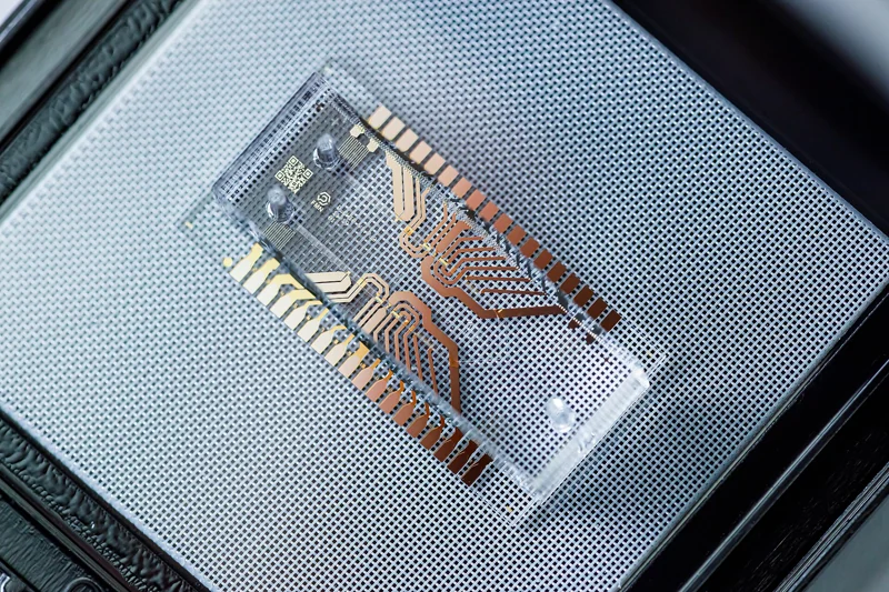
A chip may contain human cells arranged along small channels or separated by a porous support. Fluid delivery controls the arrival of nutrients, chemical signals or test compounds. Where the question requires it, a device can also provide movement, mechanical loading or contact between different tissue compartments. These are ways to make an experimental environment more specific, not independent guarantees of validity.
The Wyss organ-chip overview describes the combination of microfluidic engineering and living cell layers. [2] Neither component is sufficient on its own. A precisely controlled flow cannot correct an unsuitable cell population, and biologically relevant cells cannot correct an unrecognized leak. The measurement has to be interpreted as a result from the complete system, including materials, culture conditions and instrument behaviour.
A barrier experiment provides a concrete example
Imagine that the question is whether a tissue layer limits movement of a particular tracer. Researchers introduce a known amount on one side and measure its appearance on the other. The result depends on the tissue, the support, exposed area, concentration difference and time. Without those details, calling one chip “leakier” than another can conceal several different comparisons.
An educational calculation shows the importance of area. If two devices transfer the same total amount per hour but one exposes twice the tissue area, their transfer per unit area differs by a factor of two. The invented example is not a biological result. It illustrates why a useful transport measurement must specify what has been normalized and what remains different between experimental conditions.
Controls distinguish a tissue response from a device effect
A test compound might bind to tubing, change the fluorescence of a tracer or damage cells. Each could alter an assay without producing the intended biological mechanism. An appropriate control targets an alternative explanation: a device without tissue can reveal some material effects, an independent detector can test readout interference, and a cell-survival assay can identify widespread injury.
Positive controls are useful too. If a system fails to respond to a change known to affect the chosen function, a negative result with a new compound is hard to interpret. No single list of controls fits every experiment. The principle is to identify the most plausible ways the result could arise and design comparisons that separate them. That often matters more than adding another cell type.
Cell source affects what the result represents
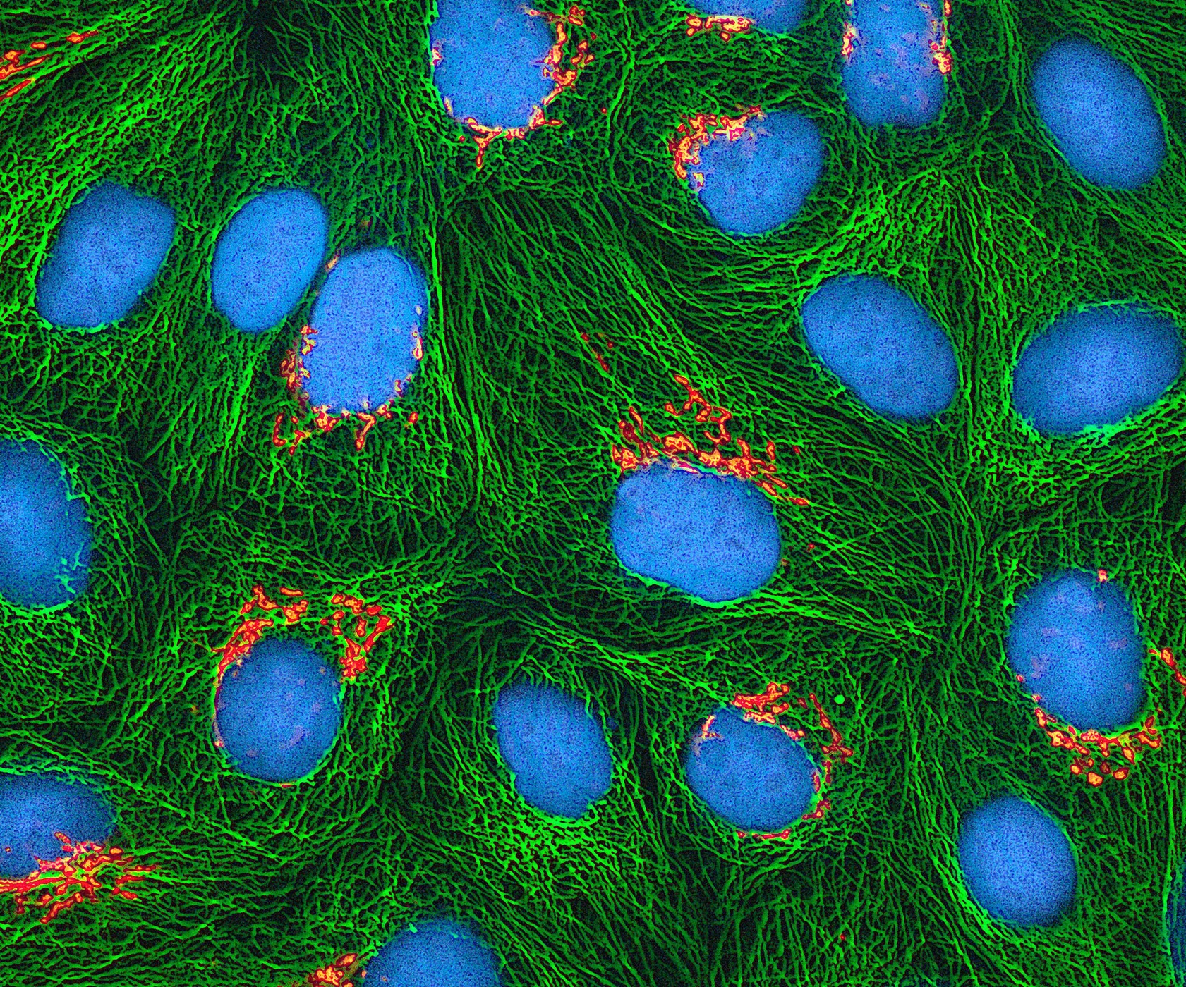
Primary cells obtained from tissue can retain useful donor-related features, while established cell lines offer different advantages in consistency and availability. Stem-cell-derived models introduce opportunities to investigate particular genetic backgrounds, alongside questions about maturation and differentiation. The appropriate source depends on the function being measured and the decision the model is intended to support.
Donor diversity also has to be represented honestly. Ten devices made from one donor can help characterize device variation, but they do not establish the range of responses among ten people. A study may need independent donors, repeated preparations and repeated devices within each preparation. Keeping those levels separate makes the strength and limits of the evidence easier to understand.
A recent colon-chip study shows the value of reconstruction
The 2026 human IBD-on-a-chip paper used patient-derived intestinal epithelial cells and stromal fibroblasts to investigate disease-associated behaviour. Recombining tissue components helped the researchers examine which interactions contributed to measured responses. [3] This is a good example of complexity added for a reason: separate cell populations create comparisons that a single mixed culture cannot provide.
It also illustrates why the question should stay close to the experiment. Evidence about a tissue interaction does not establish a complete prediction of disease progression or treatment response in a person. Additional biological systems, independent replication and clinical evidence are needed for those broader claims. The model's strength lies in controlled access to part of the mechanism.
The nominal dose may differ from the tissue exposure
A researcher may prepare a known concentration at the inlet, but the cells can experience a different time-dependent exposure. A substance may bind to device materials, be taken up by cells or change chemically before reaching the measurement region. Fluid movement also determines how quickly a concentration change arrives and how long it lasts.
This distinction matters when comparing devices or connecting tissue compartments. The same inlet label does not guarantee the same exposure at the tissue. Where the question requires it, measuring the substance in relevant streams helps connect the intended dose with the biological response. The chip's chemical behaviour is part of the model, just as its cell types and geometry are.
Validation depends on the intended use
A model can perform well for one task and poorly for another. Predicting whether a compound disrupts a particular barrier is different from predicting its full safety profile. Validation should therefore specify the input, the measurement, the comparison set and the decision being supported. Known examples are useful only if they meaningfully challenge the proposed use.
For instance, a model intended to distinguish harmful from harmless exposures should be assessed with both kinds under a predefined interpretation rule. Testing only striking positive examples can make performance appear stronger than it is. Reproducing the work in another laboratory adds evidence that the result is not dependent on a single operator or instrument. These demands turn a promising model into a tool whose limitations can be described quantitatively.
Linking chips increases both possibility and uncertainty
Connecting tissue models can help investigate how a substance changed in one compartment affects another. It also raises practical questions about circulating medium, relative tissue size, timing and the interpretation of concentration. An arrangement of several human cell types is not automatically a model of whole-body exposure. The connections themselves need to be justified.
The public benefit could be more informative experiments, better selection of candidates for further research and reduced reliance on unsuitable models. Those gains should be demonstrated for specific uses. Organ chips are most persuasive when the design, controls and measured function form a coherent answer to a defined question. Their contribution is experimental access to relationships that would otherwise be difficult to separate, rather than a promise to reproduce every feature of human life.
A model earns confidence through validation against the job it is meant to do.
Sources & further reading
Numbered references connect claims to their source. The main text is our original explanation; conceptual examples are identified as imagined.
- NIH NCATS — Tissue Chip for Drug ScreeningGovernment research programme · tissue-model development
- Wyss Institute — Human Organs-on-ChipsResearch institution · technology background
- Özkan et al. — Human inflammatory bowel disease-on-a-chipResearch paper · Nature Biomedical Engineering · 21 May 2026
Source review: 30 Oct 2026. Editorial standards.