A capture problem becomes an electrical design problem
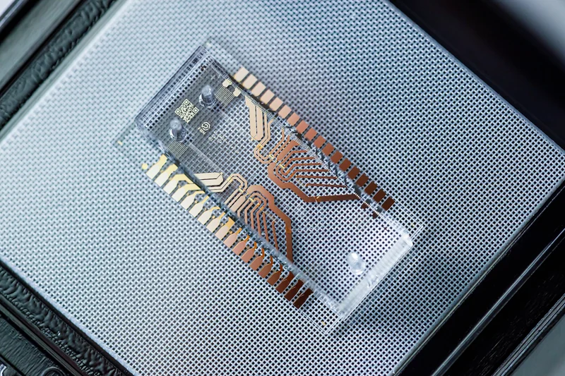
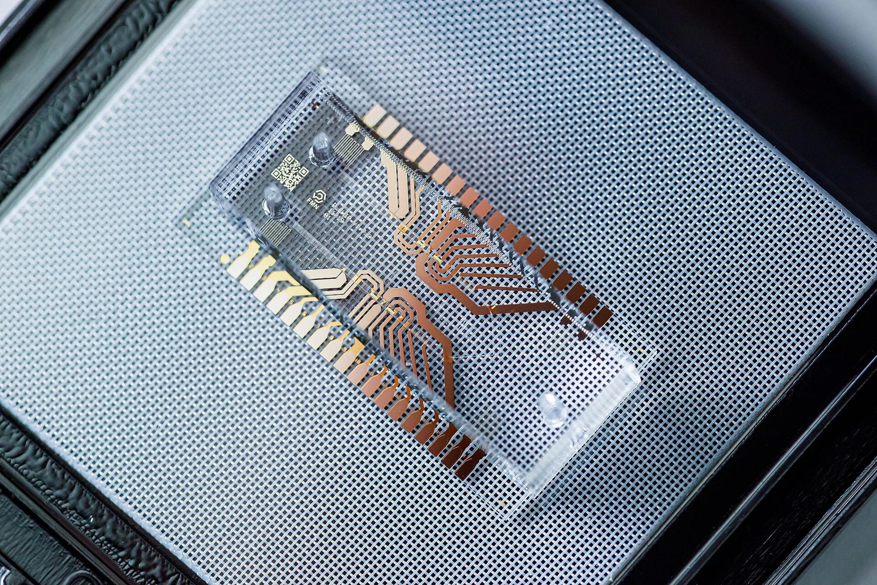
A cell entering a microchannel does not automatically settle into a position where its contents can be measured. Flow carries it forward, gravity and cell properties affect where it travels, and a later reaction needs a small, isolated space. Microwells provide such spaces, but passive wells can leave empty positions or collect more than one cell. A 2 November 2026 study in Microsystems & Nanoengineering tests whether a patterned electric field can guide cells into microwells etched directly into glass.
The researchers combine the wells with negative dielectrophoresis, or nDEP. Dielectrophoresis moves polarizable particles in a non-uniform electric field. With nDEP, the effective force points away from the field maximum; the ring-shaped electrode in this device creates a lower-field region at its center. The well is placed at that center, so the field geometry helps direct a cell into a physical compartment. The paper reports tests with polystyrene beads and HEK-293 cells, then demonstrates in-well lysis. [1]
Why negative DEP can matter in salty biological buffers
The choice between positive and negative DEP depends on how the cell polarizes relative to the surrounding liquid at the applied frequency. Positive DEP attracts particles toward stronger fields; negative DEP repels them from those regions. The direction is therefore not a label for “good” or “bad” capture. It is a way to place the useful force where the device needs it, given the cell and medium properties.
The authors present compatibility with higher-conductivity, physiological media as a motivation for nDEP. That matters because buffer conductivity changes the electrical environment and can make some electrode configurations difficult to use. A 2024 DEP platform study likewise describes frequency-dependent changes in cell behavior and emphasizes that electrode layout and operating conditions shape manipulation. These are design considerations, not a guarantee that every nDEP setup will work in every biological buffer. [1] [3]
Etch the well where the field already has a useful minimum
The key geometry is a glass microwell etched at the center of a ring electrode. That placement addresses a constraint: the modeled downward nDEP capture region is vertically limited, so simply adding a raised or layered trapping structure could put the cell outside the most useful part of the field. Simulations were used to study electrode spacing, ring diameter, electrode width, channel height and cell size before the team selected a fabrication geometry.
The fabricated wells averaged about 23.7 micrometres deep, with a bottom diameter near 21.8 micrometres. The channel height was set at 25 micrometres. These measurements help explain the engineering tradeoff: a well must be large enough to receive a cell but placed close enough to the electrode plane for the field to exert the intended force. The design was tested at the scale of individual cells, not as a finished cartridge for routine sampling. [1]
Capture, counting and lysis are separate demonstrations
The experiments use alternating-current excitation at megahertz frequencies and flow through the chip. The paper reports capture observations for beads and cells, with the biological cells requiring stronger excitation than the more uniform plastic beads. This difference is expected to matter: beads offer a more consistent size and dielectric response, whereas cells vary in both. Success with a bead standard is therefore a useful engineering check, but it cannot substitute for testing biological samples.
The platform also records impedance changes as material enters the capture region. With a calibration curve and a consistent buffer, that electrical signal can help estimate how many targets were captured; the authors note that optical monitoring remains another option. They then sealed wells with oil and used freeze-thaw treatment to lyse captured cells. Each stage answers a different question: can the field position objects, can an electrical signal track occupancy, and can an isolated reaction occur in the well? [1]
A worked example separates occupancy from a single-cell result
Imagine 100 wells under a microscope after a capture run: 70 contain one cell, 10 contain multiple cells and 20 are empty. The single-cell occupancy is 70 of 100 wells, or 70 percent. If a bulk impedance signal changes by an amount calibrated against beads, it may estimate total captured material, but it cannot by itself tell which wells hold one cell versus two. Image-based occupancy and impedance are complementary measurements, not interchangeable definitions of capture efficiency.
This is a teaching example, not the study’s reported result. A rigorous comparison would state the denominator, distinguish single from multiple occupancy, and show how cells were counted across repeated runs. It would also test different cell types and sample conditions. The paper uses impedance analysis and microscopy as evidence for its device concept, but those measurements do not establish a universal performance rate for patient samples. [1]
The platform adds a compartment, not a complete assay
A microwell can keep a captured cell and its reaction products spatially separated during processing. That is valuable for experiments where the identity of an individual cell matters. Yet cell lysis is only a starting operation: sequencing, protein measurement or electrophysiology would each need their own chemistry, controls, detection limits and interpretation rules. The authors identify such applications as possibilities for further work, rather than outcomes already demonstrated by this device.
The distinction resembles a broader lesson from DEP engineering. A 2024 study of ADEPT showed cell trapping, separation and interactions using an adaptable electrode system; it also describes the electronics, sample handling and application choices that sit around the force itself. Together, these studies show why “on-chip” is not one performance claim. A working field pattern, an isolated cell, a dependable assay and a useful biological conclusion are successive validation steps. [3]
What remains to be measured
The new study is a proof of concept. The authors identify practical constraints, including off-target capture outside the wells, cell loss when displacing oil at higher flow rates, and a voltage range where cell viability must be protected. They also call for further electrode refinement and parameter optimization. These limitations are important because stronger forces or faster flow may improve one part of the workflow while damaging another.
Next studies could report replicated capture distributions across cell types, quantify the share of empty and multi-cell wells, and compare impedance estimates with direct images under changing buffer conductivity. They could also test whether each captured cell remains viable after electrical exposure and whether lysate can be recovered reproducibly for a named downstream assay. Until such evidence exists, the result supports a promising microdevice architecture—not a validated clinical diagnostic or a broadly interchangeable single-cell platform. [1]
A useful principle for microfluidic analysis
The study’s central contribution is the alignment of three scales: an electric-field minimum, a glass microwell and a single-cell reaction space. That alignment offers a design route for researchers who need both active capture and a place to process the captured object. It also makes the engineering problem easier to reason about: field distribution guides motion, geometry retains the cell, and a separate measurement tests what happened.
For analytical chemistry and life science, the practical value will depend on matching those parts to the intended sample and readout. nDEP changes where the force points; etched microwells change where a cell can remain; impedance offers one route to monitoring occupancy. The study makes a coherent case for testing these elements together, while its own limits clarify what still needs independent measurement.
The work integrates active nDEP capture with etched microwell compartments and demonstrates impedance monitoring and cell lysis. It remains a research-stage device concept with cell-type, operating-condition and downstream-assay validation still required.
Sources & further reading
Numbered references connect claims to their source. The main text is our original explanation; conceptual examples are identified as imagined.
- Jiang et al. — Microwell-based nDEP multifunctional microfluidic system for single-cell capture and analysisPrimary research paper · Microsystems & Nanoengineering · 2 November 2026
- PubMed — Microwell-based nDEP multifunctional microfluidic system for single-cell capture and analysisNLM bibliographic record and abstract · 2 November 2026
- Julius et al. — Portable dielectrophoresis for biology: ADEPT facilitates cell trapping, separation, and interactionsPrimary research paper · Microsystems & Nanoengineering · 1 March 2024
Source review: 15 Nov 2026. Editorial standards.