INDEPENDENT SCIENCE KNOWLEDGE
Editorial standardsSource libraryMicroTAS 2010 archive
Molecules in Context:Research, Observation, Technology, Applications & Society — 10 Perspectives
SCIENCE IN CONTEXT / 10 PERSPECTIVES
IMAGE PROVENANCE

Scientific photography & image credits

Real microscopy and device photographs appear alongside clearly identified generated laboratory illustrations. Source, authorship and reuse terms are recorded below.

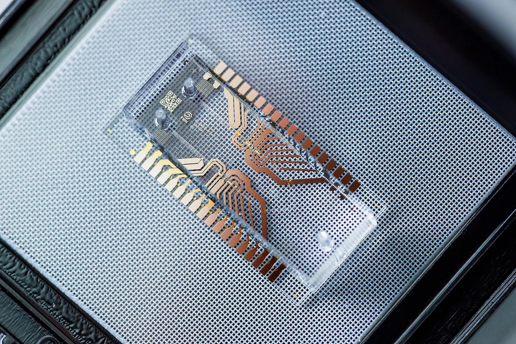
Microfluidic chip on a glass substrate, photographed against a dark background.
SCIENTIFIC PHOTOGRAPH

Microfluidic Device

A microfluidic device for reagent dilution and mixing; contextual device photograph, not the colon-chip study device.

Author: FMNLab

Source: Original image and metadata

Reuse: CC BY 4.0

Preparation: Resized and encoded as WebP; no retouching. Card layouts may crop the display.

View the full image →
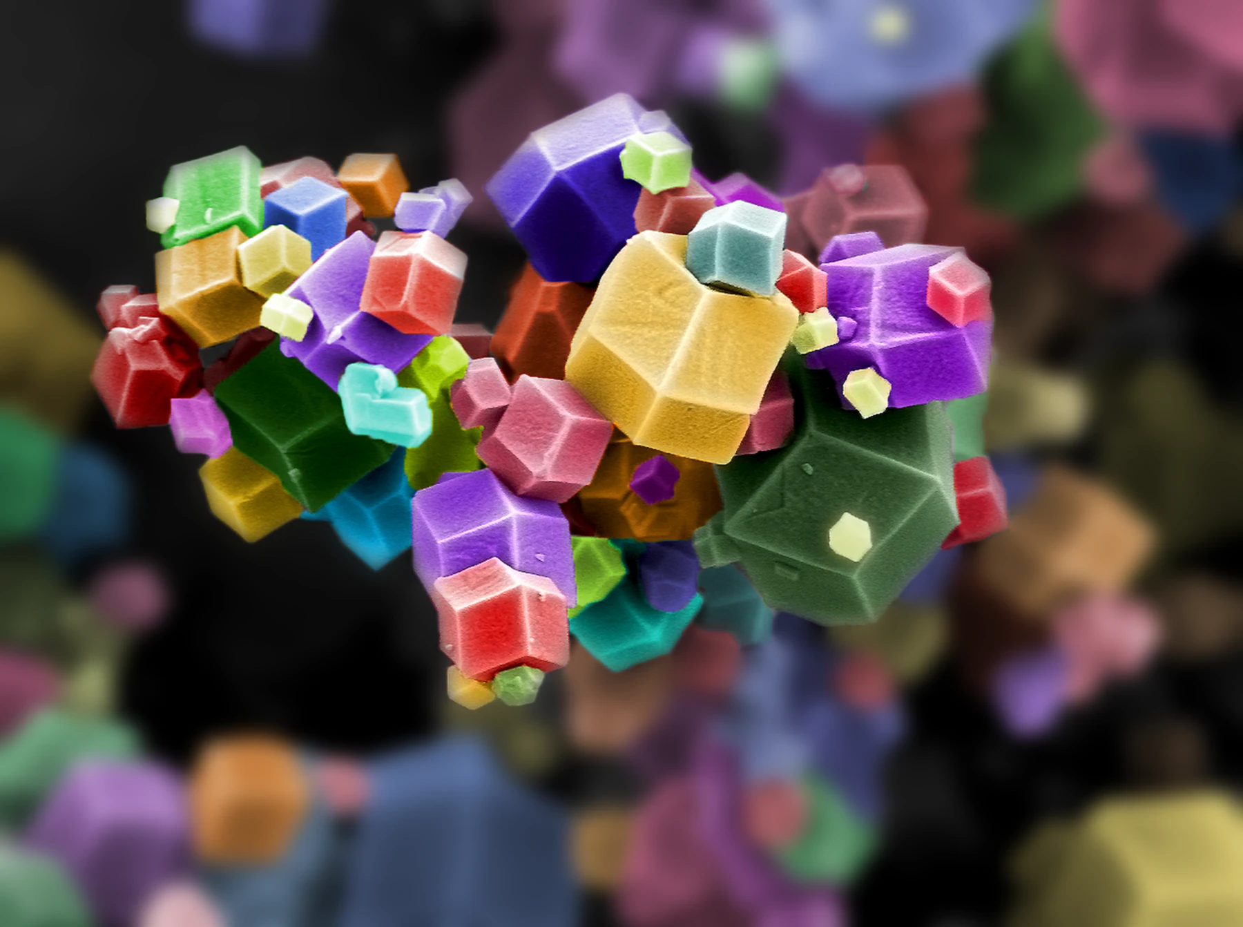
Scanning electron micrograph of dodecahedral manganese-based metal–organic framework crystals.
SCIENTIFIC PHOTOGRAPH

Mof Crystals

Colourized scanning electron micrograph of a manganese-based metal–organic framework. Colour is a visualization aid, not the material’s visible-light appearance; this is not a figure from the Nobel background document.

Author: Snemani2023

Source: Original image and metadata

Reuse: CC BY 4.0

Preparation: Resized and encoded as WebP; no retouching. Card layouts may crop the display.

View the full image →
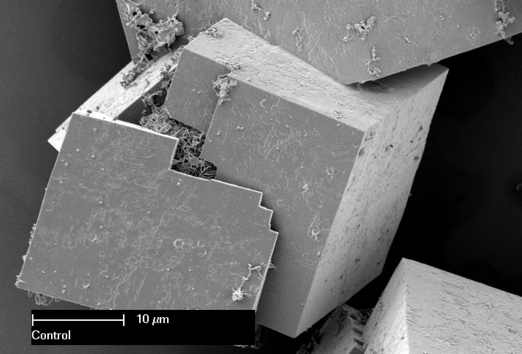
Electron microscope image showing a seed within a metal–organic framework crystal.
SCIENTIFIC PHOTOGRAPH

Mof Seed

Scanning electron micrograph of metal–organic framework crystals from CSIRO research, with the original 10 μm scale bar retained. Contextual microscopy, not a figure from the Nobel background document.

Author: Dr Paolo Falcaro and Dr Dario Buso, CSIRO

Source: Original image and metadata

Reuse: CC BY 3.0

Preparation: Resized and encoded as WebP; no retouching. Card layouts may crop the display.

View the full image →
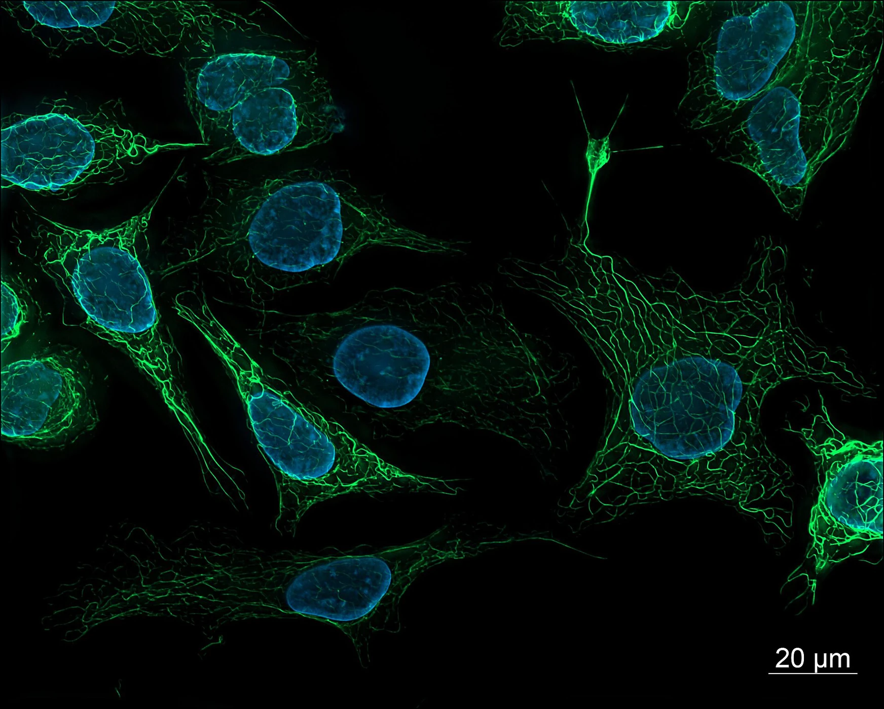
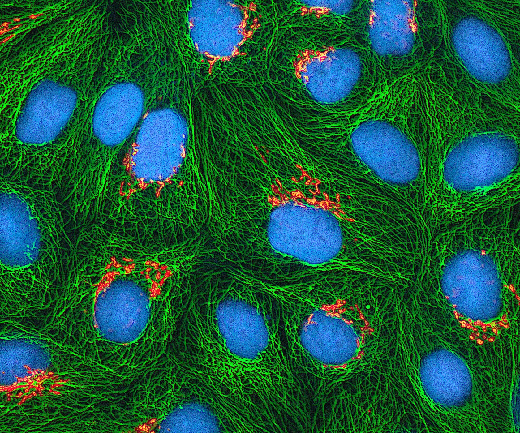
Multiphoton fluorescence micrograph of cultured HeLa cells with green microtubules, orange Golgi regions and cyan DNA.
SCIENTIFIC PHOTOGRAPH

Hela Cells

Cultured HeLa cells under multiphoton fluorescence microscopy: Golgi apparatus in orange, microtubules in green and DNA in cyan. This is a contextual cell image, not tissue from the colon-chip study.

Author: National Institutes of Health (NIH)

Source: Original image and metadata

Reuse: Public domain

Preparation: Resized and encoded as WebP; no retouching. Card layouts may crop the display.

View the full image →
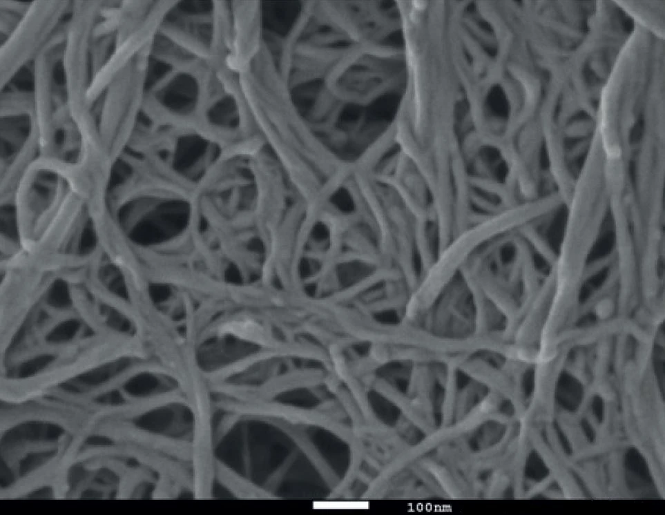
Fluorescence micrograph of human-derived cells with green vimentin filaments and blue-stained nuclei.
SCIENTIFIC PHOTOGRAPH

Human Cells

Human-derived SK8/18-2 cells: vimentin stained green and nuclei stained blue. Contextual microscopy, not a figure from an article’s cited experiment.

Author: ZEISS Microscopy from Germany

Source: Original image and metadata

Reuse: CC BY 2.0

Preparation: Resized and encoded as WebP; no retouching. Card layouts may crop the display.

View the full image →
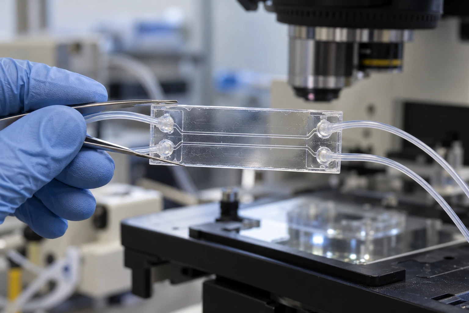
Microscope image of an interwoven cellulose nanofibre network.
SCIENTIFIC PHOTOGRAPH

Cellulose Fibres

Cellulose nanofibre network. A real microscopy image illustrating polymer structure at a small scale.

Author: Qspheroid4

Source: Original image and metadata

Reuse: CC BY-SA 4.0

Preparation: Resized and encoded as WebP; no retouching. Card layouts may crop the display.

View the full image →
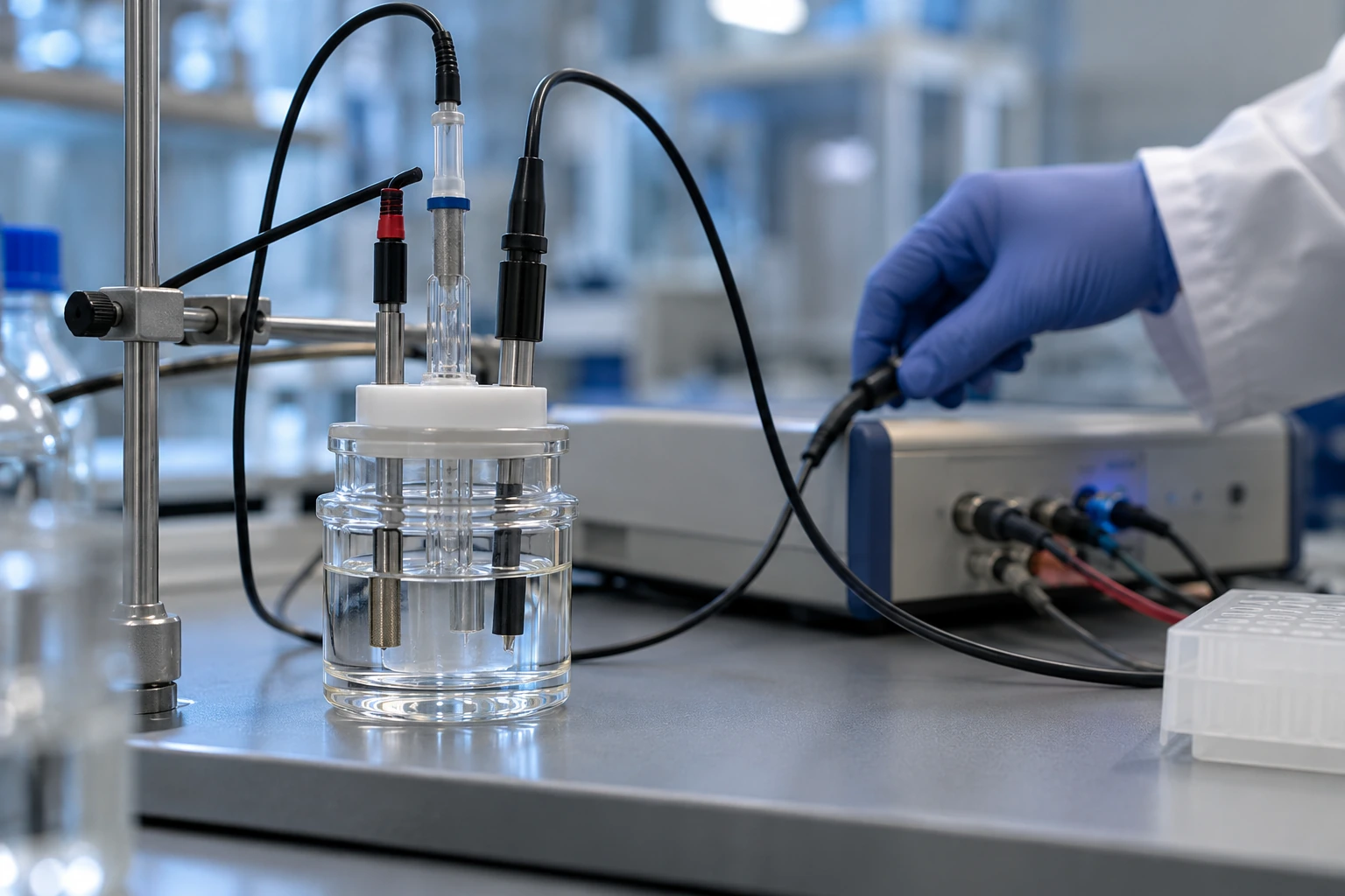
Generic microfluidic laboratory setup with a transparent device, fine tubing and a microscope.
GENERATED EDITORIAL ILLUSTRATION

Organ Chip Lab

AI-generated laboratory illustration. A generic microfluidic device near a microscope; not the device or tissue used in the colon-chip study.

Creation: Built-in image generation for Molecules in Context: Research, Observation, Technology, Applications & Society — 10 Perspectives. This is a photorealistic illustration of a generic research setting, not a photograph or an experimental result.

Preparation: Generated with the built-in image tool, then resized and encoded as WebP. Card displays may crop the frame.

View the full image →
Generic electrochemistry laboratory cell with electrodes and instrument leads.
GENERATED EDITORIAL ILLUSTRATION

Electrochemistry Lab

AI-generated laboratory illustration of an electrochemical measurement setup. It does not depict the cited PFAS experiment or its operating conditions.

Creation: Built-in image generation for Molecules in Context: Research, Observation, Technology, Applications & Society — 10 Perspectives. This is a photorealistic illustration of a generic research setting, not a photograph or an experimental result.

Preparation: Generated with the built-in image tool, then resized and encoded as WebP. Card displays may crop the frame.

View the full image →
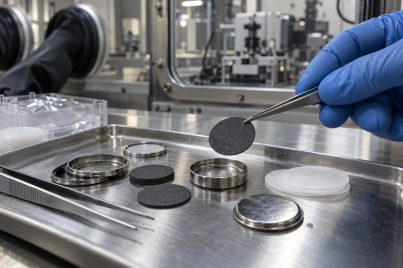
Battery materials research illustration showing coin-cell casings, electrode discs, separator membranes and precision tweezers.
GENERATED EDITORIAL ILLUSTRATION

Battery Materials Lab

AI-generated laboratory illustration of battery-material handling. It is not a photograph of a particular battery chemistry, recycling process or experimental result.

Creation: Built-in image generation for Molecules in Context: Research, Observation, Technology, Applications & Society — 10 Perspectives. This is a photorealistic illustration of a generic research setting, not a photograph or an experimental result.

Preparation: Generated with the built-in image tool, then resized and encoded as WebP. Card displays may crop the frame.

View the full image →
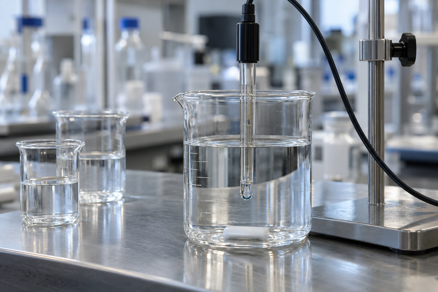
A glass pH electrode immersed in clear liquid on a laboratory bench.
GENERATED EDITORIAL ILLUSTRATION

Chemical Analysis Lab

AI-generated laboratory illustration of pH measurement in a clear solution. It does not show a measured pH, treatment performance or a real experimental result.

Creation: Built-in image generation for Molecules in Context: Research, Observation, Technology, Applications & Society — 10 Perspectives. This is a photorealistic illustration of a generic research setting, not a photograph or an experimental result.

Preparation: Generated with the built-in image tool, then resized and encoded as WebP. Card displays may crop the frame.

View the full image →
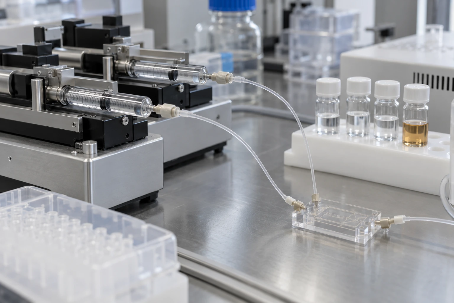
A generic formulation research setup with syringe pumps, fine tubing, a mixing chip and sample vials.
GENERATED EDITORIAL ILLUSTRATION

Rna Formulation Lab

AI-generated laboratory illustration of formulation research. The hardware is contextual, not a specified recipe or a device validated for RNA nanoparticle production.

Creation: Built-in image generation for Molecules in Context: Research, Observation, Technology, Applications & Society — 10 Perspectives. This is a photorealistic illustration of a generic research setting, not a photograph or an experimental result.

Preparation: Generated with the built-in image tool, then resized and encoded as WebP. Card displays may crop the frame.

View the full image →
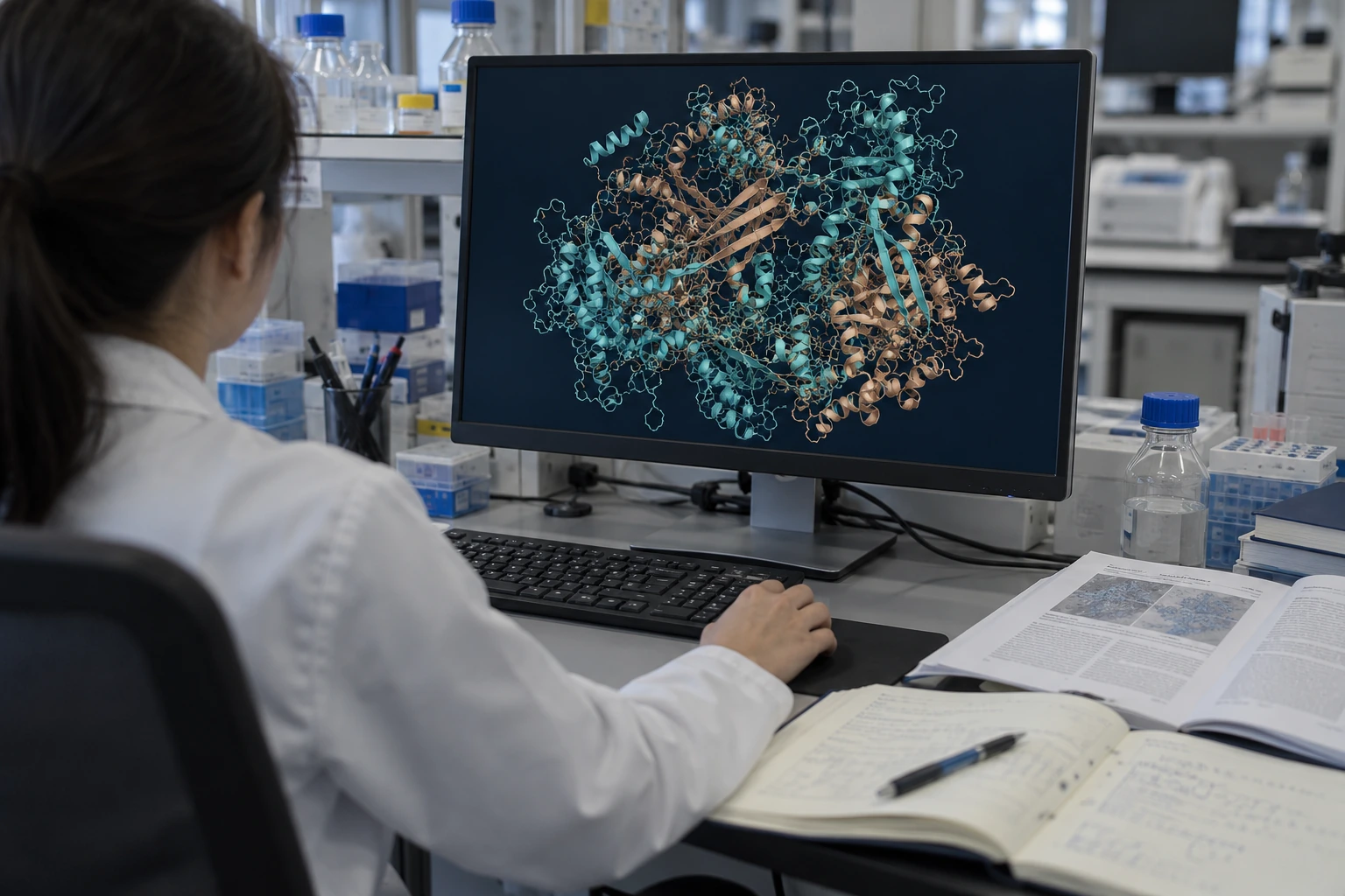
A researcher at a laboratory workstation displaying a complex protein-style molecular visualization.
GENERATED EDITORIAL ILLUSTRATION

Computational Biology Lab

AI-generated editorial illustration of computational structural biology. The displayed molecular visualization is illustrative, not a predicted structure or output from AlphaFold 3.

Creation: Built-in image generation for Molecules in Context: Research, Observation, Technology, Applications & Society — 10 Perspectives. This is a photorealistic illustration of a generic research setting, not a photograph or an experimental result.

Preparation: Generated with the built-in image tool, then resized and encoded as WebP. Card displays may crop the frame.

View the full image →