The question changes when liquid moves
A static infection assay places microbes and host cells together in a well and measures what remains attached after a chosen interval. That is useful for controlled comparisons, but the corneal surface is continually exposed to moving tear fluid. Flow can carry cells and compounds past the surface, change how long they remain near it, and impose mechanical forces. A result measured without motion therefore answers a narrower question than whether an anti-adhesion effect persists in a flowing environment.
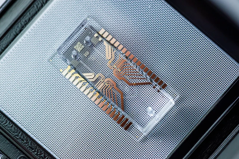
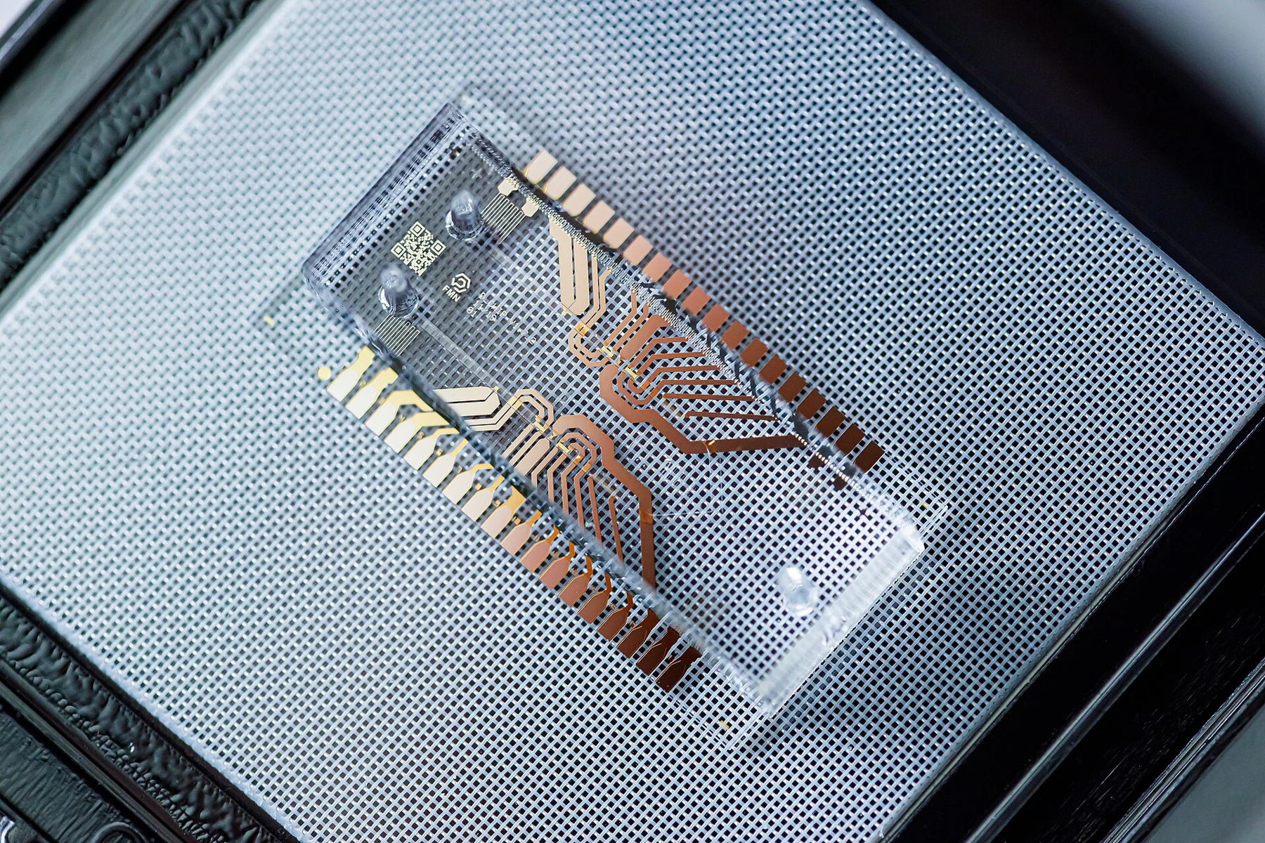
In a paper published 5 November 2026, Urwin and colleagues built a plate-based microfluidic model using a human corneal epithelial cell line, HCE2. The small test channel held a cell monolayer while a pressure-controlled system moved liquid across it. Time-lapse microscopy and automated image analysis tracked attached bacteria over two hours. The investigators compared two model strains, Staphylococcus aureus SH1000 and Pseudomonas aeruginosa PAO1, and tested pretreatment with unfractionated heparin (UFH), a compound previously proposed to interfere with bacterial attachment. [1]
A simple intervention produced different results
Under flow, UFH pretreatment substantially reduced S. aureus attachment relative to the paired untreated channel. At 60 minutes, the authors reported a 65% reduction; the difference remained statistically significant at later measured timepoints. In the same microfluidic setup, UFH pretreatment did not produce a statistically significant reduction in P. aeruginosa attachment. Yet in parallel static well-plate assays, pretreatment reduced attachment for both organisms. [1]
That pattern matters more than a single “effective” or “ineffective” label. The same pretreatment behaved differently depending on which organism was tested and whether the liquid moved. Flow did not simply weaken every measured effect: the S. aureus result persisted, while the P. aeruginosa result changed. It is a reminder that an assay condition can interact with the biology being measured, rather than acting as a neutral background.
What the microchannel contributes
The device gave the team a way to observe attachment during continued delivery of bacteria and liquid instead of measuring only an endpoint after a static incubation. Researchers could image the same cell layer repeatedly, quantify attached cells over time and compare treated and control channels. A flowing channel also makes transport explicit: bacteria arrive, pass through, contact the epithelial surface, and may remain attached or leave with the liquid. Those processes are compressed into a single final count in many static assays.
In physical terms, flow supplies a changing environment at the cell surface. The experiment does not isolate one universal “flow effect”; attachment reflects contact opportunities, surface interactions, fluid conditions and the behavior of each bacterial strain. The study’s channel and operating conditions create a controlled laboratory model, not a complete replica of blinking, tear turnover, immune activity, tissue layers or the changing chemistry of an infected eye. [1]
How far the result reaches
The finding supports a methodological conclusion: screening anti-adhesion candidates only in static conditions can miss differences that appear under flow. It does not show that UFH prevents keratitis, treats an established infection or is suitable for use in the eye. The authors examined pretreatment of cells, not a clinical dosing strategy, and the work was performed in vitro. A reduction in bacterial attachment is also not the same endpoint as killing bacteria, clearing an infection or improving vision.
The size and scope of the experiment matter. The detailed treated-versus-control comparisons used four biological repeats, two laboratory strains and a short two-hour attachment window. Statistical significance in this setup indicates evidence for a difference under these tested conditions; it does not guarantee the same result across clinical isolates, patients, other flow regimes or longer infections. PubMed independently records the paper’s publication and abstract, including the contrast between static and flow results. [2]
A useful next experiment is a better comparison
The model is most useful as a bridge between a simple screen and more complex systems. A next stage could test additional strains and clinically relevant isolates, vary flow in a controlled way, and compare dose and exposure duration while keeping the cell layer and imaging analysis consistent. Replication across independent experiments would help establish how stable the organism-specific difference is. Those are research directions, not results reported by this study.
For readers interpreting a laboratory claim, the practical question is not just whether a compound “worked.” Ask what endpoint was measured, under what transport conditions, against which organisms and with how many independent repeats. Here, the endpoint was bacterial attachment to cultured corneal epithelial cells. The mismatch between static and flowing conditions is the central result—and a reason to treat microfluidics as a way to sharpen experimental questions, not as a shortcut from a chip to a clinical conclusion.
A pressure-driven microfluidic channel let researchers watch bacteria attach to corneal epithelial cells under continuous flow. Unfractionated heparin pretreatment reduced Staphylococcus aureus attachment in this model but did not measurably reduce Pseudomonas aeruginosa attachment, even though static assays suggested reductions for both. The result illustrates how assay conditions can affect screening conclusions; it does not establish a clinical therapy.
Sources & further reading
Numbered references connect claims to their source. The main text is our original explanation; conceptual examples are identified as imagined.
- Urwin et al. — A microfluidic model of bacterial keratitis to quantify the efficacy of anti-infective compounds under flowPeer-reviewed primary research · Journal of Applied Microbiology · published 5 November 2026
- PubMed — A microfluidic model of bacterial keratitis to quantify the efficacy of anti-infective compounds under flowBibliographic record and abstract · NCBI PubMed · 2026
Source review: 21 Nov 2026. Editorial standards.