Why the intestinal barrier is a research problem
The intestine is an exchange surface that must do several jobs at once. Its lining encounters food components and microbes, absorbs useful substances and helps control what reaches the tissue beneath it. A barrier is therefore not simply a sealed wall. Its function depends on living cells, their connections, the surrounding material and signals from neighbouring tissue.
Inflammatory bowel disease, or IBD, includes Crohn's disease and ulcerative colitis. These are chronic conditions involving intestinal inflammation, with different patterns of disease. A laboratory model cannot reproduce a person's whole illness, but it can help investigate a specific interaction that is difficult to isolate in a patient. That is the motivation for bringing patient-derived cells into a carefully controlled microfluidic environment. [1]
What was reported in May 2026
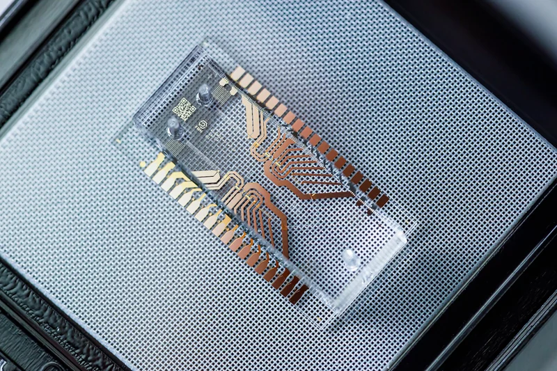
On 21 May 2026, the Wyss Institute described research published in Nature Biomedical Engineering using human colon chips constructed with patient-derived epithelial cells and stromal fibroblasts. The study examined disease-associated changes in the barrier, inflammation and fibrosis, as well as the influence of mechanical deformation and pregnancy-associated hormones. [2]
The primary paper reports experiments in which cells from healthy and diseased tissue compartments were recombined. These comparisons implicated disease-associated fibroblasts as contributors to several measured IBD-like features. This is more specific than observing that an unhealthy-looking chip resembles a disease: changing the source of one tissue component provides evidence about its role within the model. It does not establish that the same component explains every case of IBD. [3]
Two tissue compartments provide different information
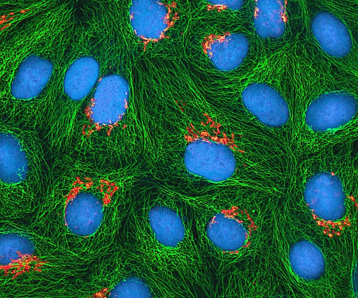
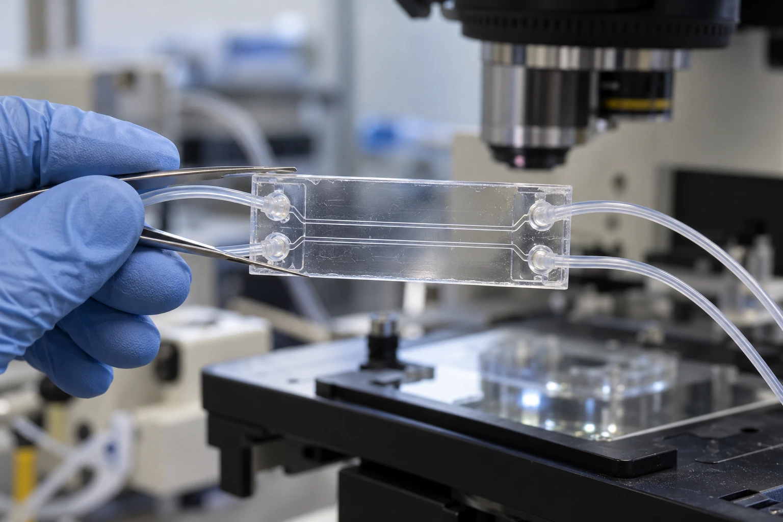
Epithelial cells form the lining facing the channel that represents the intestinal interior. Fibroblasts occupy the supporting tissue compartment. They are not just passive packing material: their interactions with the epithelium are part of the biological question. A permeable interface lets the compartments exchange molecular signals while allowing researchers to control their surroundings separately.
Separate compartments also make it possible to collect different outflows. An increase in a measured molecule on one side can be compared with changes on the other side and with changes in barrier transport. This does not identify a causal pathway by itself, but it gives an experiment more resolution than a single mixed sample. The architecture creates opportunities for distinctions that would otherwise be lost in an average.
Flow and movement are experimental variables
A static culture exposes cells to a different environment from a tissue supplied continuously with fluid. Flow can replenish nutrients, remove secreted substances and produce forces at a surface. Mechanical stretching adds another kind of stimulus. Those effects need to be distinguished because changing both at once makes it harder to identify which produced a response.
In the colon-chip study, peristalsis-like deformation was investigated alongside the tissue composition. The paper describes disease-related responses that depended on this mechanical context. [3]
For a reader, the important principle is that a device's motion is a controlled variable rather than decorative realism. If motion changes a biological measurement, the next question is whether that change is reproducible and relevant to the condition being studied.
How a barrier measurement becomes evidence
Imagine an educational experiment in which a detectable tracer is introduced into one channel. Researchers measure how quickly it appears in the neighbouring compartment. Faster passage may indicate a change in barrier transport, but several explanations remain possible: altered cell junctions, damaged tissue, changes in exposed area or a leak in the device itself.
Controls help separate those possibilities. A channel without cells characterizes the support material; microscopy can reveal gaps; a cell-survival measurement can distinguish a regulated response from widespread injury. Repeated measurements over time can show whether the change precedes or follows another event. These are general examples of experimental reasoning, not a description of every assay in the reported study. They show why one striking image is rarely enough to establish a mechanism.
Why swapping cell sources is informative
Suppose a model contains two interacting cell populations, A and B. A comparison of healthy A plus healthy B against diseased A plus diseased B changes both populations simultaneously. If the result differs, either population, their interaction or another accompanying difference could matter. Testing healthy A with diseased B, and then diseased A with healthy B, narrows the possibilities.
The approach is especially useful when cells can be compared within donor-related experimental designs. It still needs careful replication: many chips produced from one donor do not represent many independent people. Biological diversity enters at the donor level as well as the device level. The study's tissue-recombination strategy illustrates the value of controlled reconstruction, while the limited donor sampling constrains how broadly its results can be generalized. [3]
The tissue environment is more than a list of cells
Cells interact with the extracellular material around them as well as with soluble signals. Fibroblasts help produce and remodel this environment. Fibrosis involves changes in connective tissue, so measuring only a few secreted molecules can leave an important part of the tissue response unexamined.
This suggests further mechanistic questions rather than automatic conclusions. Does a changed matrix alter how forces reach the epithelial layer? Does that alter the signals cells exchange? Which change comes first? A controlled tissue model can help separate such relationships if the necessary measurements are included. The broader lesson is that cell identity, physical surroundings and time belong together when interpreting a disease model.
What the hormone experiments can and cannot say
The researchers also exposed chips to pregnancy-associated hormone conditions and measured changes in disease-related responses. This provides a way to investigate direct effects on the modelled tissue environment, separated from many other changes occurring during pregnancy. The institutional report presents this as a mechanistic research opportunity, not a treatment recommendation. [2]
Pregnancy involves interacting endocrine, immune, metabolic and circulatory changes. A controlled hormone exposure recreates only part of that setting. A chip result therefore cannot determine an individual person's likely disease course or guide changes to medication. Its value is in identifying a testable relationship that can be examined with additional models and clinical evidence. Keeping that distinction clear makes the finding useful without turning it into unsupported personal advice.
From an experimental window to public benefit
A convincing tissue model needs a defined purpose. It might help investigate a pathway, compare candidate interventions or identify a measurable sign of disease activity. Each purpose calls for different evidence. A model that helps explain one cell interaction has not automatically been validated to predict how a medicine will perform in a population.
NIH's tissue-chip programme aims to improve the bridge between laboratory discoveries and human biology. [4] Progress along that bridge requires reproducible manufacturing, consistent cell preparation and tests whose results mean the same thing across laboratories. The potential benefit is better-informed research decisions before expensive clinical work. The immediate achievement is narrower and still substantial: a controllable system in which an otherwise complicated tissue relationship can be examined directly.
A more realistic model is valuable when its extra complexity answers a specific biological question.
Sources & further reading
Numbered references connect claims to their source. The main text is our original explanation; conceptual examples are identified as imagined.
- CDC — Inflammatory bowel disease basicsGovernment · disease background
- Wyss Institute — Decoding inflammatory bowel disease on a chipResearch institution · 21 May 2026
- Özkan et al. — Human inflammatory bowel disease-on-a-chipResearch paper · Nature Biomedical Engineering · 21 May 2026
- NIH NCATS — Tissue Chip for Drug ScreeningGovernment research programme · tissue-model development
Source review: 30 Oct 2026. Editorial standards.